Glossary
A | B | C | E | F | G | H | I | K | L | M | N | O | P | R | S | T | U | V
Amblyopia: One eye does all the visual work while the other "lazy" eye develops poor vision from disuse.
Aphakia: The lens of the eye has been removed.
Astigmatism: Due to irregularities in the curvature of the cornea or lens, light rays are not focused clearly in a point on the retina. This condition results in distorted vision, especially at nearby distances.
Bilateral: This term means that both sides are affected or "two sided". Bilateral cataracts are cataracts in both eyes.
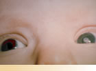
Congenital Cataract: A congenital opacity (cloudiness) in the lens of the eye is present at, or develops soon after birth.
Emmetropia: Normal vision produced when the light rays are focused clearly in a point on the retina.
FDA: The US . Food & Drug Administration is the government body that is charged with protecting the public health by assuring the safety & security of drugs, medical devices, food, cosmetics and products that produce radiation. It must also help to speed innovations that make medicines and foods more effective, safer and more affordable.
Glaucoma: A higher than normal pressure within the eye resulting from various causes.
Hyperopia: Light rays focus at a point behind the retina. The person will see better at a distance (farsightedness).
IATS: I nfant A phakia T reatment S tudy - This study in which your child is a participant.
Intraocular Lens: This tiny prescription lens, commonly known as an IOL, is placed inside the eye to replace the eye's natural lens. The replacement occurs when the natural lens is surgically removed because it is clouded (cataract).
Keratometry: This term means the measurement of the cornea.
Lensectomy: A procedure where the natural lens of the eye is removed.
Microphthalmia: This term refers to a rare disorder in which one or both eyes are abnormally small.
Myopia : Light rays focus at a point in front of the retina. The person will see better at close distances (nearsightedness)
NEI: The N ational E ye I nstitute is one of the 27 institutes of the NIH. This body conducts and supports research that helps prevent and treat eye diseases and other vision disorders. The NEI is one of the oversight bodies of the IATS study.
NIH: The N ational I nstitutes of H ealth is the steward of medical and behavioral research for the USA . It funds and oversees scientific studies at universities and research institutions across the nation and is comprised of 27 institutes and centers.
Ophthalmologist: This medical doctor (MD) or doctor of osteopathy (DO) specializes in the medical and surgical care of the eyes and visual system and in the prevention of eye disease and injury.
Optometrist: This doctor of optometry (OD) is not a medical doctor. An optometrist can diagnose vision problems & eye diseases, prescribe eyeglasses, contact lenses and drugs to treat eye disorders, but cannot perform surgery.
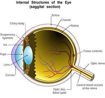
This diagram is used by permission of Dr. Anil Rao, Metropolitan State College of Denver, Department of Biology
Cornea: The clear tissue that covers the front of the eye is sometimes compared to a watch crystal. This part of the eye is where light first enters. Normally when the cornea is touched it causes a blinking reflex in both eyes. A contact lens either completely or partially covers the cornea.
Iris: The iris is the colored part of the eye located between the lens and the cornea. The iris controls the size of the pupil which is located in the middle of the iris.
Pupil: The opening in the middle of the iris is called the pupil. Normally the pupil is round in shape, dark in color, and equal in size to the other pupil. The pupil controls the amount of light the retina receives by becoming smaller or larger.
Ciliary body: This group of muscles controls the thickness of the lens.
Lens: The clear lens is behind the iris. Light enters the eye and reaches the lens. The lens is elastic like a rubber band. By changing shape it controls how light is focused on the retina.
Retina: The retina, which is the innermost layer of the eye, changes the light impulses received from the lens into electrical impulses. These electrical impulses are sent to the optic nerve.
Optic Nerve: The optic nerve carries the electrical impulses to the brain.
Optic Disc: The nerve head of the optic nerve is the optic disc. Since this part of the retina does not have any light sensitive cells, the optic disc is called the blind spot.
Choroid: The middle layer of the eyeball containing many blood vessels.
Pseudophakia: This term refers to the presence of an implanted intraocular lens after the removal of a natural lens with a cataract.
Randomized Clinical Trial: A randomized clinical trial designed so that study participants get a particular intervention or treatment in random order or by chance (as in the flip of a coin).
Refraction: This refers to eye testing to determine the amount of vision or visual acuity.
Rigid Gas Permeable contact lens (GP or RGP): This type of lens is custom made from firm, durable, breathable (transmits oxygen) plastic.
Silsoft contact lens: This type of lens is made from a gel like plastic which contains varying amounts of water.
Strabismus: This condition, commonly known as "crossed eyes", occurs when a person cannot align both eyes at the same time...one or both eyes may turn in, out, up or down
Tonometry: A standard test performed to determine the fluid pressure inside the eye (IOP). The intraocular pressure (IOP) is measured using a tonometer.
Unilateral: This term means that only one side is affected or "one sided". Unilateral cataract is a cataract in only one eye.
Visual Acuity Assessment: This assessment is to test the child's 'sharpness of vision'.
A | B | C | E | F | G | H | I | K | L | M | N | O | P | R | S | T | U | V